Chromaturia refers to an abnormal discoloration of urine, which can range from red, brown, orange, green, or even blue hues. It is not a disease itself but a clinical sign that may indicate underlying pathology, dietary influences, medication effects, or metabolic disorders. Diagnosing chromaturia requires a structured clinical and laboratory approach to determine the exact cause and rule out serious conditions such as hematuria, liver disease, or infections.
Below is a comprehensive overview of the tests used to diagnose chromaturia, along with their clinical significance.
1. Urinalysis (Routine and Microscopic Examination)
Urinalysis is the first-line diagnostic test for evaluating chromaturia. It provides critical information about urine composition.
Components
- Visual inspection Determines color, clarity, and turbidity
- Dipstick testing Detects
- Blood (hemoglobin/myoglobin)
- Bilirubin
- Urobilinogen
- Protein
- Glucose
- Nitrites and leukocytes
- Microscopic examination
- Red blood cells (RBCs)
- White blood cells (WBCs)
- Crystals
- Casts
- Bacteria
Clinical relevance
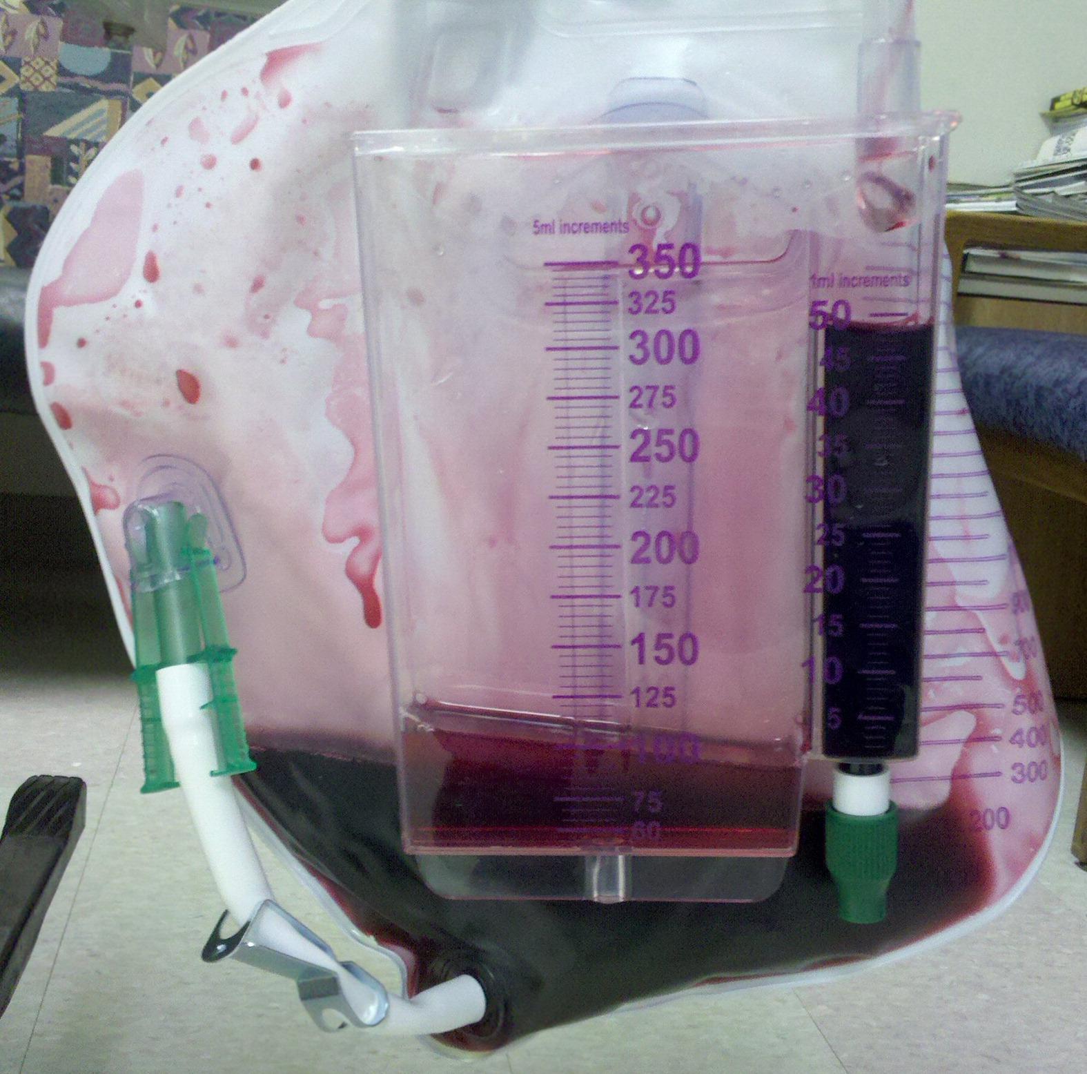
- Red or pink urine may indicate hematuria or hemoglobinuria
- Brown urine can suggest myoglobinuria or liver disease
- Green or blue urine may be linked to medications or infections
Urinalysis helps differentiate true hematuria from pigment-related discoloration.
2. Urine Culture
A urine culture is performed when infection is suspected.
Purpose:
- Identify bacterial pathogens such as Pseudomonas aeruginosa, which can cause greenish urine
- Determine antibiotic sensitivity
Indications:
- Presence of leukocytes or nitrites in urinalysis
- Symptoms like burning urination, fever, or urgency
3. Blood Tests
Blood investigations are essential to assess systemic causes of chromaturia.
Common tests
- Complete Blood Count (CBC)
- Detects infection or anemia
- Liver Function Tests (LFTs)
- Elevated bilirubin may cause dark urine
- Renal Function Tests (RFTs)
- Evaluate kidney performance
- Creatine Kinase (CK)
- Elevated in muscle breakdown (rhabdomyolysis myoglobinuria)
Clinical insight
Dark brown or tea-colored urine often correlates with bilirubinuria or myoglobinuria, which can be confirmed via blood markers.
4. Imaging Studies
Imaging helps identify structural abnormalities in the urinary tract.
Common modalities
- Ultrasound (USG):
- Detects kidney stones, tumors, cysts
- CT Scan (Computed Tomography):
- More detailed imaging for stones, masses, or trauma
- MRI (Magnetic Resonance Imaging):
- Used in complex cases
When used
- Persistent hematuria
- Suspected malignancy or obstruction
5. Cystoscopy
A cystoscopy involves direct visualization of the bladder and urethra using a thin scope.
Indications
- Unexplained hematuria
- Suspected bladder tumors
- Chronic urinary symptoms
What it detects
- Tumors
- Stones
- Inflammation
- Structural abnormalities
6. Urine Cytology
Urine cytology examines urine for abnormal or malignant cells.
Purpose
- Detect cancers of the urinary tract, especially bladder cancer
Indications
- Persistent unexplained discoloration
- High-risk patients (e.g., smokers, elderly)
7. Specialized Biochemical Tests
Certain cases of chromaturia require targeted biochemical analysis.
Examples:
- Porphyrin tests:
- Diagnose porphyria (reddish or port-wine urine)
- Methemoglobin levels:
- Associated with certain toxic exposures
- Homogentisic acid test:
- Detects alkaptonuria (urine turns dark on standing)
8. Drug and Toxin Screening
Many medications and toxins can cause urine discoloration.
Common culprits:
- Rifampicin orange urine
- Metronidazole dark urine
- Certain laxatives pink/red urine
One important antimicrobial drug is Nitazoxanide, often prescribed at nitazoxanide 500mg dosage for parasitic infections. While generally well tolerated, medications in this class may occasionally alter urine color indirectly through metabolic byproducts or liver enzyme interactions. Therefore, a medication history is crucial when evaluating chromaturia.
Screening approach
- Review prescription and over-the-counter drugs
- Toxicology tests if poisoning is suspected
9. Dietary Assessment
Food intake can significantly influence urine color.
Examples
- Beetroot red urine (beeturia)
- Carrots orange urine
- Artificial dyes various colors
Diagnostic approach
- Dietary recall
- Temporary elimination diet
This helps avoid unnecessary invasive testing when discoloration is benign.
10. Genetic and Metabolic Testing
In rare cases, chromaturia is linked to inherited metabolic disorders.
Conditions
- Alkaptonuria
- Porphyria
- Maple syrup urine disease
Tests
- Genetic screening
- Enzyme assays
- Metabolic panels
Diagnostic Approach Summary
A systematic approach ensures accurate diagnosis:
- History taking
- Duration and color of urine
- Associated symptoms
- Medication use (including nitazoxanide 500mg if relevant)
- Dietary habits
- Initial tests
- Urinalysis
- Blood tests
- Further evaluation
- Imaging
- Culture
- Cytology
- Advanced testing
- Specialized biochemical or genetic tests
Clinical Importance of Early Diagnosis
Identifying the cause of chromaturia is crucial because it can range from benign conditions (dietary causes) to serious diseases such as
- Kidney disorders
- Liver disease
- Urinary tract malignancies
- Severe infections
Delays in diagnosis may lead to complications, especially in cases involving hematuria or systemic illness.
Conclusion
Chromaturia is a visually striking but diagnostically complex clinical sign. A combination of urinalysis, blood investigations, imaging studies, and specialized tests is used to identify the underlying cause. Careful consideration of medications including treatments like nitazoxanide 500mg dietary factors, and systemic conditions is essential for accurate diagnosis.
A structured, stepwise evaluation allows clinicians to distinguish harmless causes from serious pathology, ensuring timely treatment and optimal patient outcomes.
Join our community to interact with posts!